Panoramic X-rays
Wednesday, January 18, 2012
Dental X-Rays
Dental X-Rays are pictures of the teeth , bones, and soft tissues around them to help find problems with the teeth, mouth, and jaw. X-ray pictures can show cavities, hidden dental structures (such as wisdom teeth), and bone loss that cannot be seen during a visual examination. Dental X-rays may also be done as follow-up after dental treatments

* Dental X-rays are done to :
- Find problems in the mouth such as tooth decay, damage to the bones supporting the teeth, and dental injuries (such as broken tooth roots). Dental X-rays are often done to find these problems early, before any symptoms are present .
- Find teeth that are not in the right place or do not break through the gum properly. Teeth that are too crowded to break through the gums are called impacted .
- Find cysts, solid growths (tumors), or abscesses .
- Check for the location of permanent teeth growing in the jaw in children who still have their primary (or baby) teeth .
- Plan treatment for large or extensive cavities , root canal surgery , placement of dental implants , and difficult tooth removals .
- Plan treatment of teeth that are not lined up straight (orthodontic treatment) .
Without X-rays , dentists may miss the early stages of decay between teeth .

How do dental X-rays work ?!
As X-rays pass through your mouth they are mostly absorbed by teeth and bone because these tissues, which are called hard tissues , are denser than cheeks and gums , which are called soft tissues. When X-rays strike the film or a digital sensor, an image called a radiograph is created. Radiographs allow the dentist to see hidden abnormalities, like tooth decay, infections and signs of gum disease, including changes in the bone and ligaments holding teeth in place .
Risks
Radiation used in dental X-rays is so low that there is very little chance of problems from having the X-rays .
Pregnant women may not want to have routine dental X-rays taken until after they give birth. Although there is no proof that a routine dental X-ray could harm the fetus , dentists usually suggest you wait to have your X-rays until after the baby is born. Delaying the X-ray for a few months will not result in further harm to teeth in most cases. There are times when the severity of the dental problem requires an X-ray to deal with an urgent concern .

Results
Dental X-Rays are pictures of the teeth, bones, and soft tissues around them to help find problems with the teeth, mouth, and jaw.

# Types of Dental X-Rays ;
- Intraoral X-rays are the most common type of dental X-ray taken. These X-rays provide a lot of detail and allow your dentist to find cavities, check the health of the tooth root and bone surrounding the tooth, check the status of developing teeth, and monitor the general health of your teeth and jawbone.
> Types of Intraoral X-Rays ; .
1- Bitewing X-rays .

2- Periapical X-rays .

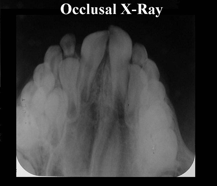
3- Occlusal X-rays .
- Extraoral X-rays show teeth, but their main focus is the jaw and skull. extraoral X-rays are used to look for impacted teeth, monitor growth and development of the jaws in relation to the teeth, and to identify potential problems between teeth and jaws and the temporomandibular joint .
> Types of Extraoral X-Rays ;
1/ Panoramic X-rays .

2/ Tomograms .
3/ Cephalometric projections .
4/ Sialography .
5/ Computed tomography .
Subscribe to:
Posts (Atom)